
Email: info@pascomedical.co.uk
Email: info@pascomedical.co.uk
Fluorescein dye, as part of slit lamp examinations is an important, low invasive tool in the identification and grading of anterior eye diseases particularly for dry eye, foreign bodies in the eye or damage to the cornea.
Once the dye is instilled onto the eye and the required activation time has passed, a blue light will be shined on to the patient’s eye on which the fluoresce emits a green light that is reflected to the observer instrument. The fluorescein dye will collect and pool in invisible areas of damage (including scratches, dips and holes) that will be detectable.
Since conjunctival and corneal staining patterns have proved to be particularly effective markers of dry eye, this is seen as one of the preferred techniques by clinicians due to low cost and ease of execution.
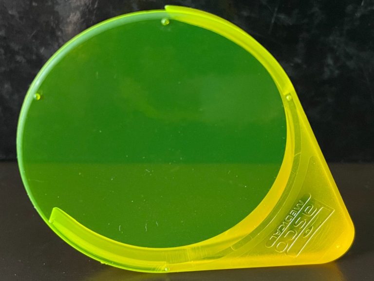
• Large lens allow maximum reflected light pass
• Once the test is complete, it can easily be removed from the slit lamp and stored for future use
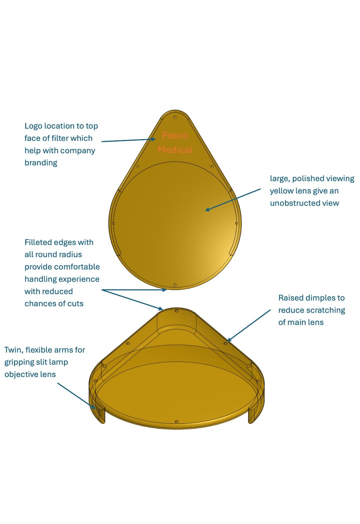
• Tear shape lens gives an unobstructed view onto the slit lamp objective lens
• Twin, flexible arms grip slit lamp objective lens tightly
• Sandblasted body reduces stray light scatter
• Logo location helps with company branding
• Raised dimples reduce scratching of main lens when filter placed on rough surface
• Edges with all round radius provide a more comfortable handling experience

Uses
A simple clip-on mechanism enables a low cost upgrade to many common slip lamps on market.
Common uses include the viewing of:
From Ophthalmologist
“It is great to see a robust, regulatory approved yellow filter based on our research, become available for all those slit lamp bio-microscopes that don’t have one built in. A yellow filter is vital for optimised viewing of fluorescein on the ocular surface and its spectral transmission needs to be carefully selected. In addition, the filter clips in place, making examination of the eye easier.”
- Prof. James Wolffsohn
Head of School of Optometry
Aston University
“The yellow filter is an amazing asset to improve the fluorescein visualisation of the cornea epithelial surface for subtle changes that may be easily missed otherwise. It is also a great teaching tool for junior ophthalmology trainees, optometrists and nurse practitioners who are starting to learn how to look for these changes as it clearly highlight them even with minimal fluorescein staining.”
- Dr Soon Ch'ng
Consultant Ophthalmologist
Birmingham and Midland Eye Centre
© 2025 Copyright. All rights reserved.
We need your consent to load the translations
We use a third-party service to translate the website content that may collect data about your activity. Please review the details in the privacy policy and accept the service to view the translations.